Soin des caries : peut-on préserver les dents ?
Les techniques actuelles de soin conduiraient trop souvent à la perte de la dent. Des spécialistes soulignent l’importance de mettre en place un modèle préventif de soins peu voire non invasifs.
La Fédération dentaire internationale a encore récemment dressé un constat alarmant sur cette maladie orale qu’est la carie dentaire, un véritable fléau mondial qui continue à sévir à un niveau endémique planétaire en dépit de la connaissance de cette pathologie et des moyens à disposition de la profession dentaire.
Face à cette situation, il est nécessaire de changer le système de prise en charge de la carie, en substituant le traditionnel modèle curatif des soins dentaires, reposant principalement sur la réalisation d’obturations dentaires qui mènent trop souvent à la perte de la dent, par un modèle de soins moins invasifs qui repose sur la préservation des tissus dentaires.
Le chirurgien-dentiste ne doit plus se borner à réparer les dents endommagées par les caries, mais à évaluer en amont le risque cariogène de chaque patient et à traiter la maladie par des mesures de prévention primaires et secondaires : application de soins dentaires a minima (scellement des sillons dentaires, reminéralisation de l’émail dentaire par l’application d’agents, micro-préparations dentaires…), en plus d’un suivi continu du patient tout au long de sa vie. En adoptant une approche médicalisée basée sur la prévention et le suivi, les patients, enfants comme adultes, pourront être épargnés par la carie.
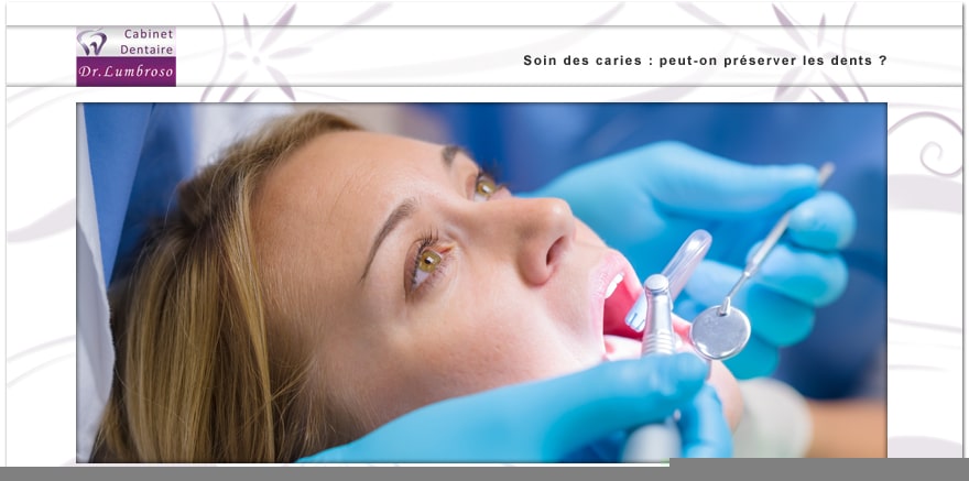
Grâce aux moyens technologiques dont disposent les praticiens, la dentisterie contemporaine permet de réaliser des soins mini-invasifs ou a minima qui restent cependant autant voire plus exigeants que des soins conventionnels. Dans ce cas, le chirurgien-dentiste doit intervenir sous anesthésie locale indolore, en posant un champ opératoire en caoutchouc sur la dent (la digue) et en s’aidant de loupes binoculaires ou de microscope dentaire pour pouvoir opérer à l’échelle de la lésion de l’émail dentaire. De plus, l’instrumentation permettant d’éliminer les tissus cariés s’est considérablement diversifiée et améliorée, à l’instar des fraises qui se sont miniaturisées et sont fabriquées dans certains cas en diamant ou en porcelaine. L’usage de ces micro-embouts diamantés abrasifs sur des instruments vibratoires standards permet par exemple d’éliminer la zone cariée de façon extrêmement précise sans détruire les tissus sains qui entourent la lésion (on parle dans ce cas de sono-abrasion).
Le praticien peut également avoir recours aux lasers oraux, davantage adaptés au traitement des tissus mous (gencive, muqueuse buccale), mais qui peuvent l’aider à compléter ou à améliorer les soins au stade précoce de la lésion carieuse. Par exemple, les lasers erbium peuvent être utilisés pour l’éviction carieuse a minima, en particulier au niveau des sillons des molaires. Le soin de la carie doit être complété avec l’emploi des composites dentaires, directement collés et polymérisés sur la dent pour obtenir une obturation étanche qui empêchera une nouvelle pénétration des bactéries cariogènes dans les tissus dentaires.
En conclusion, ces avancées technologiques n’auront d’intérêt que si le chirurgien-dentiste les combine avec un diagnostic précis et précoce des caries et un contrôle des facteurs de risques. La prise en compte de ces dimensions dans une approche globale permet de soigner efficacement la lésion carieuse tout en préservant la dent, pour le grand bénéfice des patients.
Source : le figaro.fr