History
Since the 1970s, dental implants have been a major advance in the replacement of lost teeth.
Thanks to a minimally invasive procedure, they allow the patient to smile again and chew efficiently. They are currently the most comfortable solution, and the most reliable over time (97% success rate in implantology). They avoid wearing a removable prosthesis (dental appliance) or the mutilation of healthy teeth to make a bridge (bridge between two teeth).
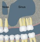
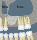
In fact, dental implants do have one constraint... In order to place an implant reliably, sufficient bone volume is required. In the upper jaw, above the molars, there is a hollow cavity in the middle of the jawbone, this "pneumatic cavity": it is the maxillary sinus. The maxillary sinus is lined with a mucous layer: this is Schneider's membrane. Very often, when the maxillary molars are extracted, the amount of residual bone under the maxillary sinus is too small to be able to place implants (at least 8 mm of bone is needed to place an implant). What can be done?
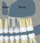
As early as the 1970s, practitioners had the idea of performing a bone augmentation under the maxillary sinus, by removing Schneider's membrane and interposing a filling material.
Today, there are two techniques for bone augmentation under the sinus:
- Elevation and partial filling of the maxillary sinus by the alveolar (or crestal) route, known as the Summers technique.
- Elevation and partial filling of the maxillary sinus using the Tatum technique or " Sinus Lift ".
These two techniques have different indications.
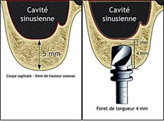
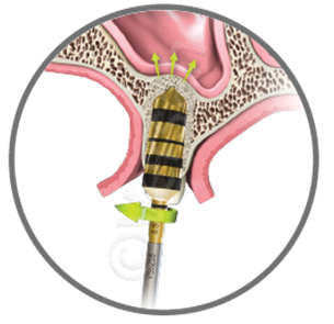
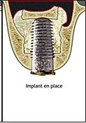
Summers' technique
The Summers technique can be used when at least 5 mm of bone remains beneath the sinus. It involves "pushing" the sinus membrane through the alveolus created for implant placement. This is now achieved by special drills that rotate in the opposite direction, which collect bone chips during drilling, condense them and gently inject them under the sinus membrane, which is then gently pushed. Depending on the volume of bone to be gained, a filling material can be interposed before the implant is placed, in the same operating time. The post-operative course is generally minor.
Sinus Lift
Le Sinus Lift est réservé aux pertes osseuses plus importantes (hauteur résiduelle < 4-5 mm). Il s’agit de réaliser une fenêtre osseuse en regard du sinus maxillaire, puis la membrane de Schneider est minutieusement décollée. Un matériau de comblement est ensuite glissé sous cette membrane, et la fenêtre est refermée. Il est parfois possible de placer les implants en même temps, mais dans la plupart des cas, il est préférable de laisser cicatriser 3-4 mois et de poser les implants après un contrôle radiographique 3D. Les suites postopératoires sont plus importantes qu’avec la technique de Summers, elles sont comparables à l’extraction de dents de sagesse incluses. Œdème et hématome surviennent parfois.
Benefits
These two meticulous operations require a large technical platform and an experienced team. This is why Dr Michaël LUMBROSO holds a University Diploma in Maxillofacial Rehabilitation Surgery(University of Paris VII), and is fully qualified to perform this type of operation. Indeed, the main risk of these operations is a tear of Schneider's membrane without the practitioner noticing it.
This is why in our practice these procedures are carried out in our operating theatre, under rigorous aseptic conditions(sterilisation chain, HEPA filter, traceability, etc...). During a Sinus Lift, in order to limit the risk of tearing the Schneider's membrane as much as possible, we use a very special instrument: the PIEZZOCHIRURGY. This instrument uses ultrasound and allows the bone window to be made without damaging the membrane underneath, as it is inactive on the soft tissue.
Moreover, in our practice, this procedure is performed under an operating microscope. This considerably increases the precision of our actions, and thus reduces the risk of tearing the membrane. And if a perforation should occur, it is almost impossible for it to go unnoticed thanks to the operating microscope. The operation is then stopped and the window closed, without any consequences for the patient's health. However, it will be necessary to intervene again after a few weeks.