When and why use Apical Microsurgery?
We perform Apical Microsurgery when traditional practice does not allow us to reach a lesion at the root tip, and when endodontic treatment and root canal filling are only partially completed, an infection may develop.
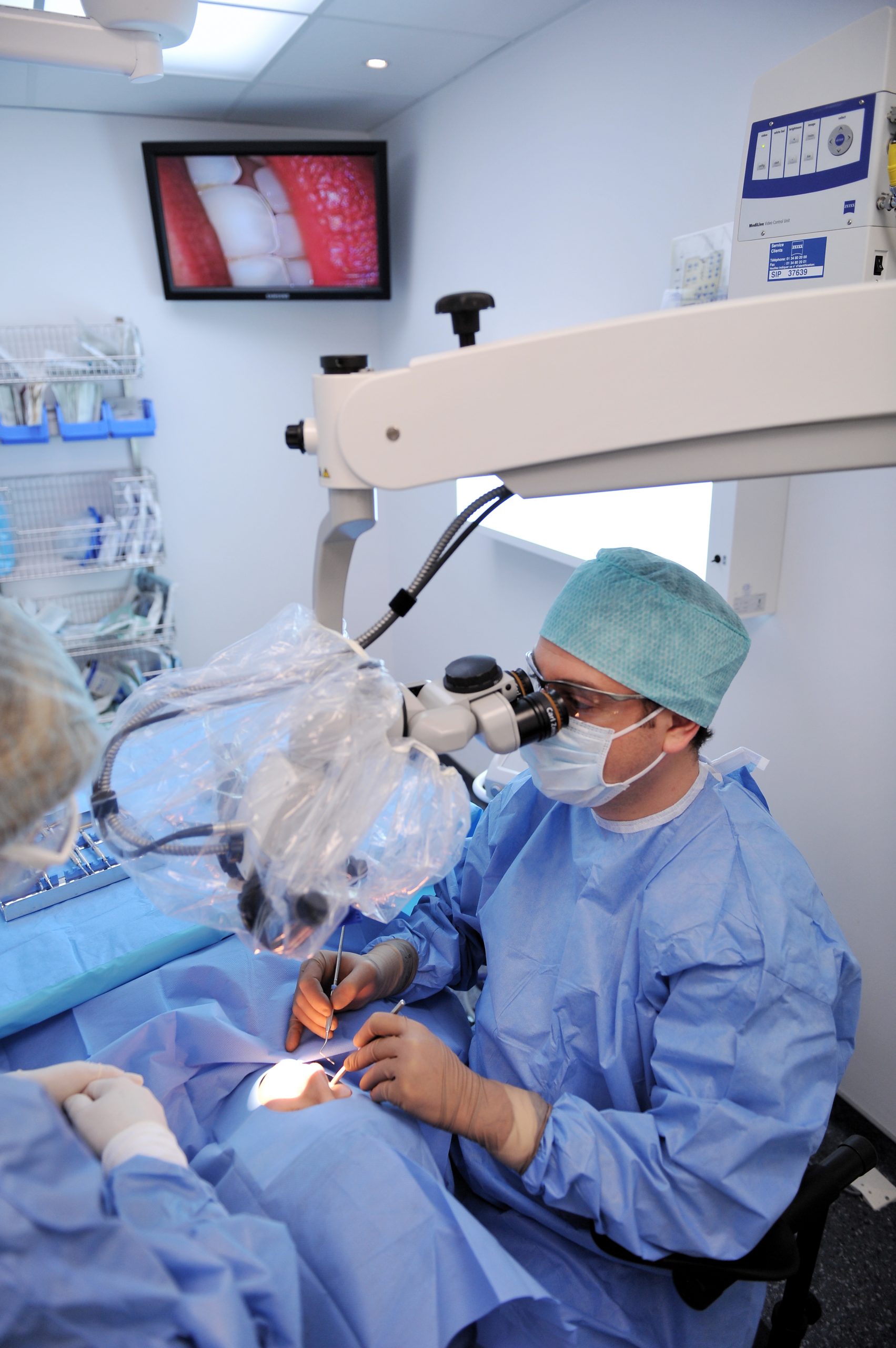
Thanks to the precision provided by the operating microscope, this operation allows a cure in a large proportion of the cases treated.
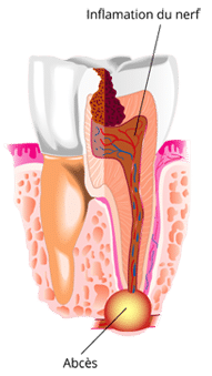
In the incompletely treated canal, the remaining pulp becomes mortified and bacteria proliferate in the unfilled portion of the canal.
A lesion(granuloma) forms at the end of the root, in the bone and ligament.
Untreated granuloma will most often progress to cyst andabscess.
The first step may be to repeat the root treatment (orthograde treatment).
When orthograde treatment does not allow the canal to be cleaned to its end, only Apical Microsurgery can remove residual bacteria.
How?
Apical resection is the surgical removal of the remaining cyst. The root tip is cut to sanitise the infected area. The bone cavity is carefully cleaned of all cystic debris.
The end of the canal is hermetically sealed, this is the retrograde filling under the operating microscope. Only the use of an operating microscope (x25 magnification) allows to work on such small surfaces.
Bone healing gradually fills the cavity left by the operation. After several months, the bone has healed and the infection has completely disappeared.
Benefits
We make precise and small incisions under a microscope. Our operations are therefore better controlled, without unnecessary damage, and the procedures performed are very precise. We thus respect the concept of "minimal surgery" and our results are all the more satisfactory.